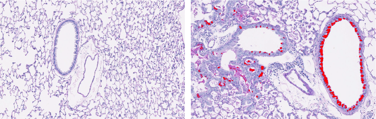
The left photo is PAS-hematoxylin staining of a section of normal lung, and the right is from a section of lung after the development of progressive lung disease long after respiratory viral infection (starting as soon as 5 days and here peaking at 7 weeks after initial infection).
In these photos, we are also using image analysis to red colorize the mucus/PAS+ material to allow for improved visualization and quantitation.
In addition, you can see the increased cellularity/hematoxylin/blue staining as a sign of inflammation and stem cell proliferation.
End result is excessive airway inflammation and mucus production and so morbidity and mortality.
Check out the published article here.